Atrioventricular (AV) block is an interruption or delay of electrical conduction from the atria to the ventricles due to conduction system abnormalities in the AV node or the His-Purkinje system. Conduction delay or block can be physiologic if the atrial rate is abnormally fast or pathologic at normal atrial rates. AV block is generally defined based on a regular atrial rhythm.
AV block is categorized as first-, second-, and third-degree AV block. First-degree AV block is defined as AV conduction slowing; on the electrocardiogram (ECG), the PR interval exceeds 0.20 seconds (sec). In second-degree AV block, some P waves conduct while others do not. This type is subdivided into Mobitz I (Wenckebach), Mobitz II, 2:1, paroxysmal, and high-grade AV block. During third-degree (“complete”) AV block, no AV conduction occurs when it should be able to occur.
Symptoms of AV block range from no symptoms to weakness, fatigue, shortness of breath, exercise intolerance, or syncope. Low cardiac output can cause hypotension and end-organ hypoperfusion. Death due to asystole is possible in complete heart block. Treatment involves correction or resolution of underlying causes and, if AV block is progressive, symptomatic, and/or persistent, a pacemaker is required.
[3]
First-degree AV block
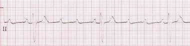
PR prolongation (PR interval >0.20 sec in adults and >0.160 sec in young children) with 1:1 AV conduction (see the image below)
A narrow QRS is associated with conduction slowing in the AV node, but it may be in the atria, and with a wide QRS, conduction slowing is in the His-Purkinje system
Marked PR interval prolongation (>0.30 sec) is usually associated with slowing in the AV node
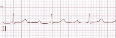 This rhythm strip shows first-degree atrioventricular block block with a PR interval of 0.360 sec. Note the fixed prolonged PR interval.
Second-degree AV block
Some, but not all, atrial impulses (generally occurring at a regular rate) failing to conduct to the ventricles
Categorized as Mobitz I, II, 2:1, paroxysmal, or high-degree AV block
Mobitz I second-degree AV block
Mobitz I second-degree AV block is associated with constant PP intervals, progressive PR prolongation, followed by one nonconducted P wave. Often, there is progressive shortening of the RR intervals during the Wenckebach cycle and a pause due to a blocked P wave that is less than the sum of two PP intervals. This form of AV block occurs in less then 50% of all types of AV block and is generally due to a block in the AV node.
[4]Rarely, it can occur with a block within or below the His Bundle; this is intra or infra-Hisian Wenckebach block, respectively.
[5]
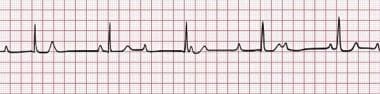
Mobitz I block rarely progresses to third-degree block. Distinguishing a block in the AV node versus infra-Hisian block can be challenging. Carotid sinus massage will worsen AV node conduction during Wenckebach block, but it may improve conduction if the block occurs below the His bundle. Exercise will worsen a block in the His-Purkinje system but improve it if the block occurs in the AV node. Mobitz I AV block usually consists of a ratio of nonconducted to conducted beats of 4:3, 3:2, and so on (see the image below).
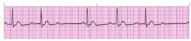 Second-degree Mobitz type I atrioventricular block. Note the prolongation of the PR interval preceding the dropped beat and the shortened PR interval following the dropped beat.
Mobitz II second-degree AV block
Mobitz II second-degree AV block is characterized by constant PP and RR intervals (see the image below). There is a constant PR interval before failure of an atrial impulse to conduct to the ventricle (small changes can occur in the PR interval after the blocked beat). The pause—including the blocked P waves—equals two PP cycles (unless an early junctional escape beat occurs). Mobitz II block occurs in the His-Purkinje system and may be associated with a narrow QRS but, as there often is in His-Purkinje disease, a wide complex QRS is commonly seen (bundle branch block) in the conducted beats. Mobitz II AV block may progress to complete AV block. Carotid sinus massage improves AV conduction in the His-Purkinje system, as it causes the sinus rate to slow; however, walking, exercise, or atropine use may lead to progressively worse AV block, as impulses may impinge upon an already damaged conduction system.
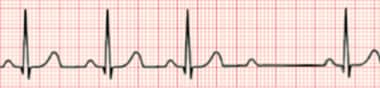 Second-degree Mobitz II atrioventricular block. Note the fixed PR interval, but after the third beat, an atrial impulse fails to conduct to the ventricle. Courtesy of Wikimedia Commons (https://commons.wikimedia.org/wiki/File:Second_degree_heart_block.png).
2:1 second-degree AV block
A 2:1 AV block is noted when every other P wave (with a regular PP interval) is conducted to the ventricle (see the image below). The block may be at the level of AV node or below. Close analysis of a long rhythm strip may help define the location of 2:1 AV block, especially during particular maneuvers. For example, if the block is in the His bundle or below, exercise, walking, or even standing may improve the AV node conduction; however, it will slow the ventricular rate and increase the level of the block. Carotid massage or atropine use may improve conduction if the block is in the His-Purkinje system, but it will worsen the block if it is at the level of the AV node.
Look for the presence of a bundle branch block. If present, a 2:1 AV block is more likely occurring in the His-Purkinje system.
 A constant PP interval and normal PR interval in conducted beats is present. This progresses to 2:1 atrioventricular (AV) block. A 2:1 AV block can be present with conduction delay in the AV node or His-Purkinje system, but it is more likely to be in the AV node for all patients (with a greater chance of AV block in the His-Purkinje system if there is a bundle branch block). Review extended monitoring strips because Mobitz I or Mobitz II may be present at other times, and this might help to determine the level of the block.
Paroxysmal AV block
Paroxysmal AV block is a form of second-degree AV block that is not persistent or repetitive, as there is an abrupt block in AV conduction. It may occur due to paroxysmal augmentation in vagal activation.
High-degree AV block
High-degree AV block consists of multiple constant P waves in a row that do not conduct. The conduction ratio can be 3:1 or higher; the PR interval of conducted beats can be constant (with His-Purkinje disease) or variable (with AV nodal block). This is distinct from complete AV block, because some P waves conduct to the ventricle. Atrial fibrillation with pauses greater than 5 seconds is due to a high-degree AV block. It can be associated with a narrow or wide QRS complex.
 High-degree atrioventricular block is demonstrated with a 4:1 atrial-to-ventricular conduction ratio. Note the P wave prior to the QRS conducts whereas the others do not. Courtesy of Life in the Fast Lane (https://lifeinthefastlane.com/ecg-library/basics/high-grade-block/), Edward J Burns, MD, Sydney, Australia.
Third-degree AV block
No atrial impulses conduct to the ventricle when they should conduct (ie, complete AV block)
The P waves, generally regular, reflect a sinus node rhythm independent from a junctional (narrow) or ventricular (wide) escape rhythm (see the image below), and they may be regular or irregular
The junctional escape rate is 40-60 bpm, and the ventricular escape rate is 20-40 bpm
Non-sinus atrial rhythms such as atrial fibrillation, atrial flutter, or atrial tachycardia can be associated with third-degree AV block
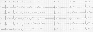
 This rhythm strip shows third-degree atrioventricular block (complete heart block). The atrial rate is faster than the ventricular rate, and no association exists between the atrial and ventricular activity. Courtesy of Life in the Fast Lane (https://lifeinthefastlane.com/ecg-library/basics/complete-heart-block/), Edward J Burns, MD, Sydney, Australia.
AV block mimickers
Blocked premature atrial complexes can be a physiologic process. These are not considered a form of AV block but are due to ectopic beats that occur when the AV node is refractory.
Paroxysmal vagal activation can occur during sleep and may be associated with sleep apnea. Other causes include cough, vomiting, urination, eating, and defecation. When AV block is present, the PR interval slows around the time of the AV block. With high vagal tone, the PR intervals will lengthen, whereas in 2:1 AV block, for example, the PR intervals are constant.
Atrial tachycardia/atrial flutter with 2:1 AV conduction is not a form of AV block. It is due physiologic impingement on AV nodal refractoriness.
AV dissociation
During AV dissociation, the atria and ventricles beat independently of each other. AV dissociation occurs when a subsidiary pacemaker in the AV node or the ventricle overtakes the sinus node for impulse initiation due to slowing of the sinus node, or it may occur when a subsidiary site beats faster than the sinus node. Complete AV block can occur with AV dissociation, but AV dissociation alone does not indicate AV block.