2019년 1월 23일 수요일

[READ CK] Lipase and Amylase

SUMMARY AND RECOMMENDATIONS
Elevations in pancreatic enzymes are not specific for acute pancreatitis. Between 11 and 13 percent of patients admitted to the hospital with non-pancreatic abdominal pain have elevated pancreatic enzymes. (See 'Epidemiology' above.)
Elevated amylase and lipase may be due to increase in pancreatic or extrapancreatic production or a decrease in clearance (table 1 and table 2). However, in some cases pancreatic enzymes can be elevated in the absence of an identifiable disease. (See 'Causes' above.)
Patients with acute pancreatitis typically have a threefold elevation of amylase and/or lipase. However, patients with acute on chronic pancreatitis, hypertriglyceridemia-induced pancreatitis, and alcoholic pancreatitis may only have elevations in lipase. Isolated elevations in amylase may be due to salivary disease, and in rare cases, due to macroamylasemia. (See 'Isolated amylase elevation' above and 'Isolated/predominant lipase elevation' above.)
The approach to the patient with abdominal pain and elevated amylase and/or lipase is based on whether the clinical presentation is consistent with acute pancreatitis. (See 'Initial approach' above.)
In patients with characteristic abdominal pain and elevation in serum lipase or amylase up to three times or greater than the upper limit of normal, no imaging is required to establish the diagnosis of acute pancreatitis. (See 'Presentation consistent with acute pancreatitis' above.)
In patients whose clinical presentation is not consistent with acute pancreatitis, we obtain a detailed clinical history that includes medications, laboratory evaluation, and abdominal imaging to determine the cause of abdominal pain and elevated pancreatic enzymes. (See 'Presentation inconsistent with acute pancreatitis' above.)
Subsequent management in patients with a negative initial evaluation depends on the presence of continued abdominal pain. (See 'Subsequent approach' above.)
In patients in whom abdominal pain has resolved, we do not pursue additional evaluation. (See 'Patients without persistent abdominal pain' above.)
Patients with persistent abdominal pain with negative initial imaging should be evaluated for other causes of abdominal pain. Repeating amylase and/or lipase in such patients is not clinically useful. Additional evaluation with endoscopic ultrasound can be helpful in the diagnosis of chronic pancreatitis, and in patients suspected of having an occult pancreatic malignancy. (See 'Patients with continued abdominal pain' above.)
[CK] Pacemaker mediated tachycardia.

A pacemaker-mediated tachycardia (PMT) can be defined as any condition in which a pacemaker paces the ventricles at rates that are inappropriately fast. [1This can be due to (1) a rate response setting that is too sensitive, (2) tracking of atrial noise (such as what may occur with electromagnetic interference), (3) inappropriate pacemaker manipulation with rate response turned on, or (4) tracking of an atrial tachyarrhythmia related to upper rate settings.

2019년 1월 22일 화요일

[CK] Lovenox dosing. 40mg sq Qday(if BMI >40, then 50 qday)

HIGH RISK>
VTE prophylaxis: Following hip or knee replacement surgery, abdominal surgery, or in medical patients with severely restricted mobility during acute illness who are at risk for thromboembolic complications. Note: Patients at risk of thromboembolic complications who undergo abdominal surgery include those with one or more of the following risk factors: age >40 years, obesity, general anesthesia lasting >30 minutes, malignancy, history of DVT or PE.

General:
Hospitalized medical patients with acute illness at moderate and high risk for VTE (including patients with active cancer):SubQ: 40 mg once daily; continue for length of hospital stay or until patient is fully ambulatory and risk of VTE has diminished (ACCP [Kahn 2012]; ASCO [Lyman 2013]; ASCO [Lyman 2015]). Note: Extended prophylaxis beyond acute hospital stay is not routinely recommended (ACCP [Kahn 2012]; Sharma 2012). 

=> MULTIPLE risk= 40 BID possible. 

--------------------------------------------------------------------------------------------------------------------------
=> BMI >40 = Dose up to 30%

VTE prophylaxis: Note: The following recommendations may be applied to all indications for VTE prophylaxis except Bariatric Surgery, which already states specific dosing based on BMI and Pregnancy, which requires individualized dosing based on thromboembolic risk.
BMI 30 to 39 kg/m2: Use standard prophylaxis dosing.
BMI ≥40 kg/m2: Increase standard prophylaxis dose by 30% (Nutescu 2009); however, the ideal dose is unknown.
=======================
VTE prophylaxis: Note: For patients assessed to be at the highest risk (eg, Caprini score >8, stroke, active cancer, multiple risk factors), many experts combine pharmacologic methods with mechanical methods or increase the dose frequency to twice daily (eg, 30 or 40 mg twice daily). Increasing the dose should always be balanced against the risk of bleeding (Malhotra 2018; Pai 2018a; Pai 2018b).
Bariatric surgery, high VTE risk (off-label use): Note: Optimal dosing strategies have not been established. Dosing regimens based on best available evidence (Birkmeyer 2012; Borkgren-Okonek 2008; Scholten 2002).
BMI ≤50 kg/m2: SubQ: 40 mg every 12 hours initiated at least 2 hours before surgery
BMI >50 kg/m2: SubQ: 60 mg every 12 hours initiated at least 2 hours before surgery


[CK] Capsule endoscopy - post ingestion(capsule -> 2hr of CLD -> 4hr full liquid -> 6hr light snack -> after completion = normal diet)

After ingesting the M2A Capsule, do not eat or drink for at least 2 hours. You may have clear liquids 2 hours after ingesting the capsule and take any medications.After 4 hours you may have a light snack. After the examination is completed and the equipment is removed, you may return to your normal diet.

OR 


Johns Hopkins:
DIET: Clear liquids after 2 hours. Full liquids (broth, coffee, orange juice and opaque drinks) and medications after 4 hours and a snack after 6 hours.


[CK] HIT? no lovenox, heparin.

Sources of heparin include the following (table 2):
LMW heparins (eg, enoxaparindalteparinnadroparintinzaparin)
Heparin flushes (eg, for arterial lines or heparin locks)
Heparin-bonded catheters
Heparin-containing medications (eg, some forms of prothrombin complex concentrates [PCCs])
Some hematopoietic stem cell (HSC) products
Some formulations of intravenous medications
Some total parenteral nutrition preparations

[CK] EP - stopcock suture technique

Three‐way stopcock suture technique for hemostasis after ablation for atrial fibrillation

[CK] Hypoglycemia - insulin adjustment. -10% down. in a week.

<hypoglycemia - adjustment>

https://www.cadth.ca/media/pdf/InsulinStartTool_e_print.pdf

60 -> 48units bid.


[CK] Troponin. Significance > 20% with elevated first Tn or >50% from small initial Tn. (more than 50% = BIG THING!)

Myocardial infarction: 3HR -> 24HR. (peak) ~ 10days. 

  • distinct rise and fall
  • typically rise 4-8 hours post onset of symptoms in MI
  • peaks at 18-24 hours
  • levels stay elevated for 10 days (allows late diagnosis of MI, may detect re-infarction with serial testing)
Advantages
  • widely available
  • high familiarity
  • rises over 4-8 hours, peaks at 10-24 hours, declines over 10 days
  • can be used to detect reinfarction
  • predicts outcome/ mortality (if negative, then low risk 30 day outcomes)
  • area under the curve of troponin elevation correlates with infarct size
  • new assays are highly sensitive
Disadvantages
  • low specificity: multiple causes of raised troponin other than infarction e.g. PE and RV strain (see above)
  • different reference ranges for different assays
  • elevated baseline troponin in renal failure
  • diagnosis of infarction often requires repeated tests leading to a delay in diagnosis
  • ‘washout’ occurs after reperfusion
  • unclear significance of raised troponin post-cardiac surgery

What is considered a “significant change” in hsTn levels? 4

  • According to the European Society of Cardiology (ESC):
    • Increase of ≥20% if first Tn elevated, or
    • Increase of ≥50% in patients with small initial Tn elevations
https://www.aliem.com/2013/09/high-sensitivity-troponin-testing/

[CK] Periprocedural anticoagulation.

https://www.mplsheart.com/wp-content/uploads/2017/07/Periprocedural%20Mgmt%20(Cath%20Lab).pdf


<?>
After Cath -> continue AC with uptrending troponin.

[CK] Bivalirudin

An initial dose of bivalirudin 0.15-0.2 mg/kg/h, <-> 0.01 in VCU !!! 

adjusted to an activated partial thromboplastin time (aPTT) of 1.5-2.5 times the baseline value, 

has been suggested

Baseline: 30 -> 2times = 60 - 80. in general. 

Afib, LVAD, etc. 

in VCU: goal 50-75

Ix. PCI, ACS, HIT, etc.(but even afib, lvad could be) 
<35 => 0.02
<50 => 0.01

(center = 60 +- 15)

>75 => -0.01
>90 => -0.02


[CK] QTc calculation.

QT ~ 1/HR.
(tachy => shorter QT)
So needs to be adjusted.
60-100: Bazett's method is ok. (<60: too low, >100: too high => lateralized)
<60 or > 100: Framingham or something else.

QTc prolongation(definition): male > 450, female > 470
1.See II or V5 or any lead(definite one) => 1 good lead with definite T-wave
(cf. bad T-wave = small U, flat, abnormal shape etc. But if U >1mm, should be included for calc.)
2.calculate successive(2-3 beats) and obtain maximum QTc.


2019년 1월 9일 수요일

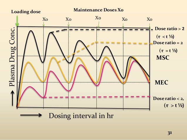
[[CK] Drug loading and maintenance graph.

loading maintenance half life graph에 대한 이미지 검색결과

[CK] contrast allergy - pre + post(we're still giving it)

The half-life in normal renal function is 30 to 60 minutes but is delayed to 20 to 140 hours in patients with severe renal function impairment.

13hr
7hr
1hr
-----prednisone medication.
1hr
-----benadryl

=> no other meds after procedure. 

[CK] OHT infection workup and treatment


Fevers and GPC chains bacteremia. Source to be indentified, but worried this is  from biliary tract. Liver US unremarkable. UA  is bening desite mentioning urinary frequency. He last performed CIC a week ago. REnal US unremarkable.
Dental work 3 weeks ago, risk for infectious endocarditis as well.
Feeling bette r on abx.

Rec:
1. Cont meropene  IV while GPCs are identified- susceptiblities released
2.  Cont Vanc  IV  while GPCs identified.
3. If remains febrile or having more positive cultures, switch to  Dapto IV to cover VRE. Hold off for now.
4. If bili  is going up, consider  repepeat MRCP and involve GI.
5. Obtain CMV PCR  in blood.
6. OBtain blood cultures follow up.
7. Cont monitoring CBC, CMP for antimicrobial toxicity.

IVF!

PAN CULTURE + GI viral + CMV/EBV PCR.
CMV/EBV IgG/IgM/DNA quantity.  + IgG level( for IVIG infusion ! )
Fungitell(Candida, Aspergillus); Platelia(Aspergillus Ag)


* Strep for sore throat.
* BK = KIDNEY Transplant.
* Legionella urine Ag. = URINE/ABDOMEN
*  respiratory/GI direct pathogen test.   = RESP

IV VANC+ZOSYN or MEROPENEM
(Dapto? for VRE but not initial)
Bil = USG + MRCP

low threshold to obtain CT (for OHT, LVAD !!!! = more likely to get!)

[CK] IDA high demand supply(IV 300 for 3 days!? Uptodate)

- IV iron sucrose 300mg daily x 3 days
- Continue po iron supplement and Ascorbic acid

Link;
https://www.uptodate.com/contents/treatment-of-iron-deficiency-anemia-in-adults?search=ida%20treatment&source=search_result&selectedTitle=1~150&usage_type=default&display_rank=1


[CK] Immunosuppressant TIP! Plus increased prednisone + hydrocortisone 50 q6h + C.DIFF!

- Restart po Vanc 125 mg every 6 hrs in the setting of recent C.Diff and restart of IV ABX
- Continue immunosuppression:
-Tacrolimus 1 mg q 12hrs - Tac level is 5.0 today
-Continue increased Prednisone 5 mg daily
-Ordered Stress dose steroid, Hydrocortisone 50 mg every 6 hours

stress dose steroid regimen에 대한 이미지 검색결과

[Q] cardiogenic shock with hypotension first??? DOBUTAMINE 5 vs DOPAMIN 5? [ ] uptodate. link

[ ] LINK ;
https://www.uptodate.com/contents/use-of-vasopressors-and-inotropes#H20


CARDIOGENIC SHOCK => DOBUTAMINE 3mcg!!!

if combination with sepsis => NE + DOBUTAMINE as goal.
                                                 May consider dopamine instead of dobutamine(more than 5mcg for
                                                 avoiding vasodilatation)!
=> BUT before decision, should consult CICU for central line placement for NE!


Dobutamine 3mcg (chronic hypotension.. slow progression)
Dobutamine is most frequently used in severe, medically refractory heart failure and cardiogenic shock and should not be routinely used in sepsis because of the risk of hypotension. Dobutamine does not selectively vasodilate the renal vascular bed, as dopamine does at low doses. (See "Inotropic agents in heart failure with reduced ejection fraction".)

vs

Dopamine 5mcg = more likely supporting Cardiac. (in urgent)
Low-dose: 1 to 5 mcg/kg/minute, results in increased renal blood flow and urine output. 
Intermediate-dose: 5 to 10 mcg/kg/minute, results in increased renal blood flow, heart rate, cardiac contractility, and cardiac output
High-dose: >10 mcg/kg/minute, alpha-adrenergic effects begin to predominate, resulting in vasoconstriction, increased blood pressure, in addition to increased heart rate, cardiac contractility, and cardiac output due to beta-adrenergic effects.


[CK] Which inotropes? inotropic agent . dobutamine vs milrinone.


- Start dobutamine 3 mcg/kg/min (BP on arrival was in 80s, probably too low for Milrinone)
- hold carvedilol
- watch on telemetry for ventricular ectopy. If high PVC burden, may need amiodarone or switch to Milrinone with addition of betablocker

2019년 1월 8일 화요일

[CK] hematuria.- bladder scan for residual and possible manual irrigation.

possible manual irrigation. =====> JUST FOR AVOIDING RETENTION!(CLOGGING) 
for

- continue to trend UOP and bladder scan pt if she is unable to void or passing small volume
- continue to trend Hgb and Cr
- if hemoglobin downtrending or hematuria worsens or pt unable to void,please contact urology as pt may require further interventions such as manual bladder irrigation 
- if approprriate continue to hold warfarin
- urine cytology
- need cystoscopy and possible MRU/CTU as an outpatient pending renal function
- pt discussed with Dr. Guruli

UPTODATE>
Initial management — The initial management of HC varies based on condition severity:
For patients with mild HC (ie, grade 1 (table 2), including those with gross hematuria without clots, adequate bladder emptying (as assessed by postvoid residual), and lower urinary tract symptoms, conservative measures, including hydration and anticholinergic bladder medications (as needed for bladder spasms), may be used in an outpatient setting after urinalysis and urine culture to exclude infection [83].
For patients presenting with the passage of blood clots (moderate or severe HC), initial management includes assessment of hemodynamic stability, hydration (or transfusion if needed), and ensuring adequate bladder drainage. Typically, in these scenarios, patients are admitted to the hospital for observation and interventions. (See 'Tempo and location of the evaluation'above.)
A large-bore (≥22 Fr) three-way Foley catheter is placed to allow for manual clot irrigation with saline. (See "Placement and management of urinary bladder catheters in adults", section on 'Specialized catheters'.)
If the urine clears following manual irrigation, subsequent conservative management with hydration alone may be sufficient. If the hematuria or clots persist despite manual catheter irrigation, CBI with normal saline may be initiated. Ensuring adequate fluid outflow from the three-way catheter while on CBI is important because if the outflow channel becomes blocked, the bladder will continue to distend with the fluid being instilled, which risks perforation. Notably, outflow obstruction can present as worsening abdominal pain or persistent bladder spasms.
If the three-way catheter has repeated obstruction, the clots cannot be removed by hand irrigation, or the patient does not improve with saline CBI, the next step is cystoscopy under anesthesia. While there are no data on the time to resolution with saline CBI alone, without repeated obstruction, we typically utilize this strategy for two to four days if the above conditions are not met and the patient's hemodynamic parameters allow. Proceeding with cystoscopy under anesthesia allows for clot evacuation (picture 1), fulguration of bleeding vessels (if they are identified), and management of any bladder tumors (if present). Notably, given the typical diffuse mucosal bleeding in HC, identifying a discrete bleeding vessel is not common. In one report of 33 patients with HC associated with cyclophosphamide or RT, 14 (42 percent) had resolution of hematuria after a single cystoscopy under anesthesia including clot evacuation or fulguration [84]. Only 4 of 11 patients who initially did not respond had resolution after a total of two or more cystoscopies.
Traditionally, cystoscopic bladder fulguration has been accomplished with electrocautery; however, some small case series note successful use of lasers for coagulation [85,86]. In these studies, a GreenLight potassium titanyl phosphate (KTP) laser (530 nm wavelength) and a 980 nm diode laser were used to manage radiation-induced HC. In an additional study, a GreenLight Xcelerated Performance System (XPS) laser (1064 nm wavelength) was successfully used in four patients who had previously failed fulguration with electrocautery [87]. Notably, attention must be paid to the depth of laser penetration and the risk of bladder perforation, as intestinal perforation and subsequent death following laser coagulation for radiation cystitis in a female patient have been reported [88].

2019년 1월 7일 월요일

[CK] [] Amyloid AL with Sotalol? Tolerate or not?

 Continue to hold Sotalol for Hx Afib: Not tolerated in AL amyloid with cardiac involvment